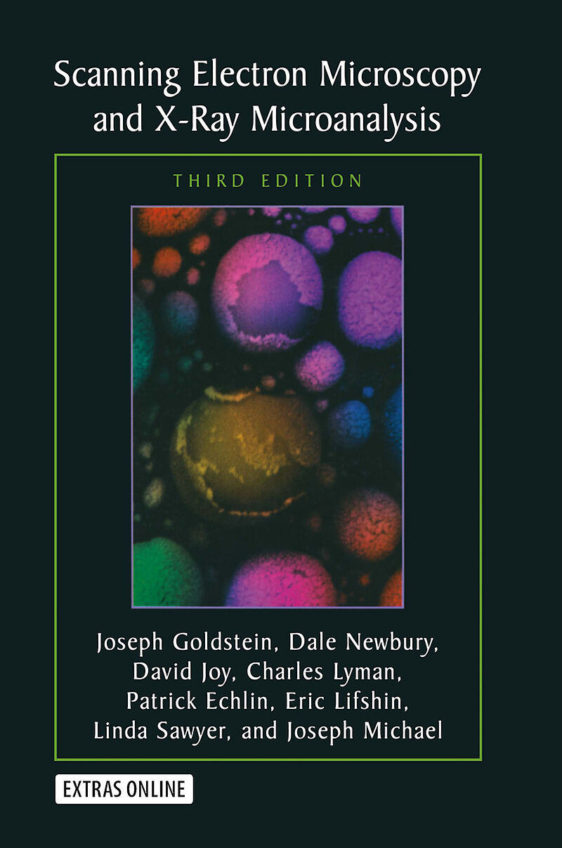
Scanning Electron Microscopy and X-Ray Microanalysis
Einband:
Fester Einband
EAN:
9780306472923
Untertitel:
Third Edition
Genre:
Allgemeines & Lexika
Autor:
Joseph Goldstein, Dale E. Newbury, David C. Joy, Charles E. Lyman, Patrick Echlin, Eric Lifshin, Linda Sawyer, J.R. Michael
Herausgeber:
Springer Nature EN
Auflage:
3. Auflage
Anzahl Seiten:
689
Erscheinungsdatum:
30.04.2007
ISBN:
978-0-306-47292-3
An ideal text for students as well as practitioners, this is a comprehensive introduction to the field of scanning electron microscopy (SEM) and X-ray microanalysis. The authors emphasize the practical aspects of the techniques described.
In the decade since the publication of the second edition of Scanning Electron Microscopy and X-Ray Microanalysis, there has been a great expansion in the capabilities of the basic scanning electron microscope (SEM) and the x-ray spectrometers. The emergence of the variab- pressure/environmental SEM has enabled the observation of samples c- taining water or other liquids or vapor and has allowed for an entirely new class of dynamic experiments, that of direct observation of che- cal reactions in situ. Critical advances in electron detector technology and computer-aided analysis have enabled structural (crystallographic) analysis of specimens at the micrometer scale through electron backscatter diffr- tion (EBSD). Low-voltage operation below 5 kV has improved x-ray spatial resolution by more than an order of magnitude and provided an effective route to minimizing sample charging. High-resolution imaging has cont- ued to develop with a more thorough understanding of how secondary el- trons are generated. The ?eld emission gun SEM, with its high brightness, advanced electron optics, which minimizes lens aberrations to yield an - fective nanometer-scale beam, and through-the-lens detector to enhance the measurement of primary-beam-excited secondary electrons, has made high-resolution imaging the rule rather than the exception. Methods of x-ray analysis have evolved allowing for better measurement of specimens with complex morphology: multiple thin layers of different compositions, and rough specimens and particles. Digital mapping has transformed classic x-ray area scanning, a purely qualitative technique, into fully quantitative compositional mapping.
The text has been used in educating over 3,000 students at the Lehigh SEM short course as well as thousands of undergraduate and graduate students at universities in every corner of the globe The authors have made extensive changes to the text and figures in this edition as a result of their experience in teaching the various concepts of SEM and x-ray microanalysis
Autorentext
This text is written by a team of authors associated with SEM and X-ray Microanalysis Courses presented as part of the Lehigh University Microscopy Summer School. Several of the authors have participated in this activity for more than 30 years.
Klappentext
This text provides students as well as practitioners (engineers, technicians, physical and biological scientists, clinicians, and technical managers) with a comprehensive introduction to the field of scanning electron microscopy (SEM) and X-ray microanalysis. The authors emphasize the practical aspects of the techniques described. Topics discussed include user-controlled functions of scanning electron microscopes and x-ray spectrometers, the characteristics of electron beam - specimen interactions, image formation and interpretation, the use of x-rays for qualitative and quantitative analysis and the methodology for structural analysis using electron back-scatter diffraction. SEM sample preparation methods for hard materials, polymers, and biological specimens are covered in separate chapters. In addition techniques for the elimination of charging in non-conducting specimens are detailed. A data base of useful parameters for SEM and X-ray micro-analysis calculations and enhancements to the text chapters are available on an accompanying CD.
Inhalt
1. Introduction.- 1.1. Imaging Capabilities.- 1.2. Structure Analysis.- 1.3. Elemental Analysis.- 1.4. Summary and Outline of This Book.- Appendix A. Overview of Scanning Electron Microscopy.- Appendix B. Overview of Electron Probe X-Ray Microanalysis.- References.- 2. The SEM and Its Modes of Operation.- 2.1. How the SEM Works.- 2.1.1. Functions of the SEM Subsystems.- 2.1.1.1. Electron Gun and Lenses Produce a Small Electron Beam.- 2.1.1.2. Deflection System Controls Magnification.- 2.1.1.3. Electron Detector Collects the Signal.- 2.1.1.4. Camera or Computer Records the Image.- 2.1.1.5. Operator Controls.- 2.1.2. SEM Imaging Modes.- 2.1.2.1. Resolution Mode.- 2.1.2.2. High-Current Mode.- 2.1.2.3. Depth-of-Focus Mode.- 2.1.2.4. Low-Voltage Mode.- 2.1.3. Why Learn about Electron Optics?.- 2.2. Electron Guns.- 2.2.1. Tungsten Hairpin Electron Guns.- 2.2.1.1. Filament.- 2.2.1.2. Grid Cap.- 2.2.1.3. Anode.- 2.2.1.4. Emission Current and Beam Current.- 2.2.1.5. Operator Control of the Electron Gun.- 2.2.2. Electron Gun Characteristics.- 2.2.2.1. Electron Emission Current.- 2.2.2.2. Brightness.- 2.2.2.3. Lifetime.- 2.2.2.4. Source Size, Energy Spread, Beam Stability.- 2.2.2.5. Improved Electron Gun Characteristics.- 2.2.3. Lanthanum Hexaboride (LaB6) Electron Guns.- 2.2.3.1. Introduction.- 2.2.3.2. Operation of the LaB6 Source.- 2.2.4. Field Emission Electron Guns.- 2.3. Electron Lenses.- 2.3.1. Making the Beam Smaller.- 2.3.1.1. Electron Focusing.- 2.3.1.2. Demagnification of the Beam.- 2.3.2. Lenses in SEMs.- 2.3.2.1. Condenser Lenses.- 2.3.2.2. Objective Lenses.- 2.3.2.3. Real and Virtual Objective Apertures.- 2.3.3. Operator Control of SEM Lenses.- 2.3.3.1. Effect of Aperture Size.- 2.3.3.2. Effect of Working Distance.- 2.3.3.3. Effect of Condenser Lens Strength.- 2.3.4. Gaussian Probe Diameter.- 2.3.5. Lens Aberrations.- 2.3.5.1. Spherical Aberration.- 2.3.5.2. Aperture Diffraction.- 2.3.5.3. Chromatic Aberration.- 2.3.5.4. Astigmatism.- 2.3.5.5. Aberrations in the Objective Lens.- 2.4. Electron Probe Diameter versus Electron Probe Current.- 2.4.1. Calculation of dmin and imax.- 2.4.1.1. Minimum Probe Size.- 2.4.1.2. Minimum Probe Size at 10-30 kV.- 2.4.1.3. Maximum Probe Current at 10-30 kV.- 2.4.1.4. Low-Voltage Operation.- 2.4.1.5. Graphical Summary.- 2.4.2. Performance in the SEM Modes.- 2.4.2.1. Resolution Mode.- 2.4.2.2. High-Current Mode.- 2.4.2.3. Depth-of-Focus Mode.- 2.4.2.4. Low-Voltage SEM.- 2.4.2.5. Environmental Barriers to High-Resolution Imaging.- References.- 3. Electron BeamSpecimen Interactions.- 3.1. The Story So Far.- 3.2. The Beam Enters the Specimen.- 3.3. The Interaction Volume.- 3.3.1. Visualizing the Interaction Volume.- 3.3.2. Simulating the Interaction Volume.- 3.3.3. Influence of Beam and Specimen Parameters on the Interaction Volume.- 3.3.3.1. Influence of Beam Energy on the Interaction Volume.- 3.3.3.2. Influence of Atomic Number on the Interaction Volume.- 3.3.3.3. Influence of Specimen Surface Tilt on the Interaction Volume.- 3.3.4. Electron Range: A Simple Measure of the Interaction Volume.- 3.3.4.1. Introduction.- 3.3.4.2. The Electron Range at Low Beam Energy.- 3.4. Imaging Signals from the Interaction Volume.- 3.4.1. Backscattered Electrons.- 3.4.1.1. Atomic Number Dependence of BSE.- 3.4.1.2. Beam Energy Dependence of BSE.- 3.4.1.3. Tilt Dependence of BSE.- 3.4.1.4. Angular Distribution of BSE.- 3.4.1.5. Energy Distribution of BSE.- 3.4.1.6. Lateral Spatial Distribution of BSE.- 3.4.1.7. Sampling Depth of BSE.- 3.4.2. Secondary Electrons.- 3.4.2.1. Definition and Origin of SE.- 3.4.2.2. SE Yield with Primary Beam Energy.- 3.4.2.3. SE Energy Distribution.- 3.4.2.4. Range and Escape Depth of SE.- 3.4.2.5. Relative Contributions of SE1 and SE2.- 3.4.2.6. Specimen Composition Dependence of SE.- 3.4.2.7. Specimen Tilt Dependence of SE.- 3.4.2.8. Angular Distribution of SE.- References.- 4. Image Formation and Interpretation.- 4.1. The Story So Far.- 4.2. The Basic SEM Imaging Process.- 4.2.1. Scanning Action.- 4.2.2. Image Construction (Mapping).- 4.2.2.1. Line Scans.- 4.2.2.2. Image (Area) Scanning.- 4.2.2.3. Digital Imaging: Collection and Display.- 4.2.3. Magnification.- 4.2.4. Picture Element (Pixel) Size.- 4.2.5. Low-Magnification Operation.- 4.2.6. Depth o…

Leider konnten wir für diesen Artikel keine Preise ermitteln ...
billigbuch.ch sucht jetzt für Sie die besten Angebote ...
Die aktuellen Verkaufspreise von 6 Onlineshops werden in Realtime abgefragt.
Sie können das gewünschte Produkt anschliessend direkt beim Anbieter Ihrer Wahl bestellen.
Loading...
Die aktuellen Verkaufspreise von 6 Onlineshops werden in Realtime abgefragt.
Sie können das gewünschte Produkt anschliessend direkt beim Anbieter Ihrer Wahl bestellen.
| # | Onlineshop | Preis CHF | Versand CHF | Total CHF | ||
|---|---|---|---|---|---|---|
| 1 | Seller | 0.00 | 0.00 | 0.00 |
